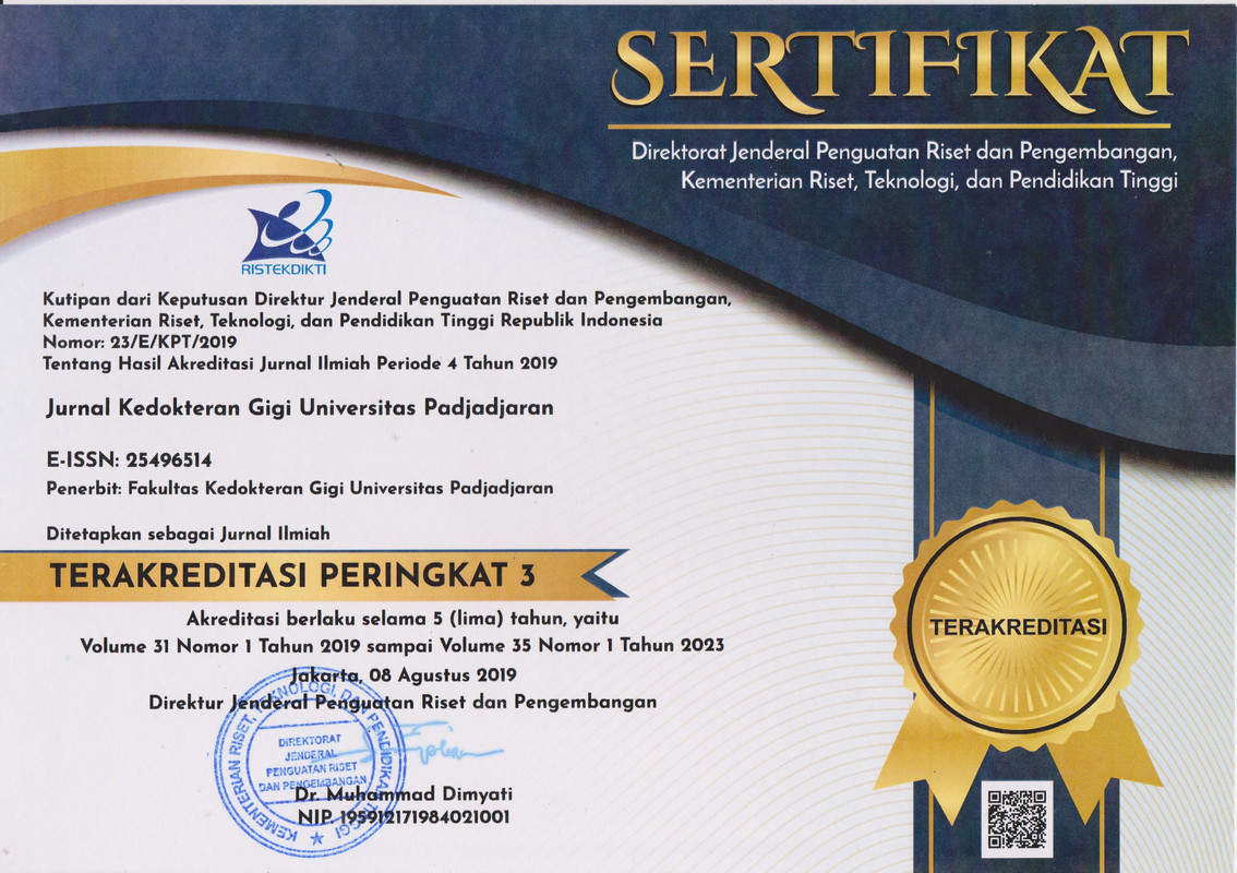
Penatalaksanaan non-bedah kasus previously initiated therapy gigi molar pertama maksila dengan lesi periapikal dan konfigurasi dua saluran akar mesiobukal
Non-surgical management of previously initiated therapy of the maxillary first molar with periapical lesions and two mesiobuccal canals configurationAbstract
ABSTRAK
Pendahuluan: Penatalaksanaan kasus penyakit pulpa yang disertai dengan lesi periapikal dapat dilakukan dengan perawatan saluran akar tanpa intervensi bedah. Preparasi biomekanis pada perawatan endodontik non-bedah dapat mengeliminasi bakteri dari saluran akar dan mencegah terjadinya infeksi berulang setelah dilakukannya obturasi. Kompleksitas anatomi menyebabkan keterbatasan preparasi biomekanis saluran akar, sehingga perawatan dapat mengalami kegagalan. Variasi anatomi saluran akar pada gigi molar pertama rahang atas umumnya terdapat pada bagian palatal akar mesiobukal yang disebut sebagai saluran akar mesiobukal kedua dengan insidensi 94%. Saluran akar mesiobukal kedua berukuran lebih sempit dan dangkal dibandingkan saluran akar mesiobukal pertama, sehingga dibutuhkan beberapa metode untuk menentukan lokasi orifis tersebut. Tujuan laporan kasus ini adalah membahas mengenai penatalaksanaan non-bedah gigi pasca perawatan endodontik parsial yang disertai dengan lesi periapikal. Laporan kasus: Pasien perempuan berusia 28 tahun datang dengan keluhan gigi belakang kiri atas sakit berdenyut sejak 1 bulan, gigi tersebut pernah dirawat saluran akarnya beberapa tahun sebelumnya. Pemeriksaan klinis menunjukkan terdapat tambalan permanen pada gigi 26. Pemeriksaan radiografis menunjukkan adanya gambaran radiopak pada email mesio-oklusal hingga mencapai dasar kamar pulpa, pelebaran ligamen periodontal, terputusnya lamina dura, serta lesi periapikal pada akar mesial dan palatal. Preparasi akses dilakukan pada gigi 26, dilanjutkan dengan penentuan lokasi orifis mesiobukal kedua. Empat saluran akar dipreparasi menggunakan instrumen rotary nickel titanium dengan teknik crown down dan dilakukan obturasi teknik kondensasi lateral. Restorasi definitif berupa mahkota penuh porselen. Simpulan: Lesi periapikal sembuh setelah dilakukan preparasi pada saluran akar mesiobukal kedua sehingga pengetahuan mengenai anatomi dan variasi internal saluran akar gigi sangat penting dalam keberhasilan perawatan endodontik non-bedah.
Kata kunci: Perawatan endodontik non-bedah; previously initiated therapy; lesi periapical; mesiobukal kedua
ABSTRACT
Introduction: The management of pulp disease cases accompanied by periapical lesions can be done by root canal treatment without surgical intervention. Biomechanical preparations for non-surgical endodontic treatment can eliminate bacteria from the root canal and prevent re-infection after obturation. However, the complexity of the anatomy limits the biomechanical preparation of the root canal so that treatment can fail. Anatomical variations of the root canal in the maxillary first molars are generally found in the palatal part of the mesiobuccal root, which is the second mesiobuccal root canal with an incidence of 94%. The second mesiobuccal root canal is narrower and shallower than the first mesiobuccal root canal, so several methods are needed to determine the location of the orifice. The purpose of this case report was to discuss the non-surgical management of teeth after partial endodontic treatment accompanied by periapical lesions. Case report: A 28-year-old female patient complained of throbbing pain in the left upper back tooth for one month. The tooth had had its root canal treated several years before. Clinical examination revealed permanent fillings on tooth 26. Radiographic examination showed the radiopaque appearance of mesio-occlusal enamel to the floor of the pulp chamber, widening of the periodontal ligament, rupture of the lamina dura, and periapical lesions of the mesial and palatal roots. Access preparation was performed on tooth 26, followed by the determination of the location of the second mesiobuccal orifice. Four root canals were prepared using a rotary nickel titanium instrument with a crown down technique and obturation with lateral condensation technique. The definitive restoration is a full porcelain crown. Conclusions: Periapical lesions healed after preparation of the second mesiobuccal root canal. This result proves that knowledge of the anatomy and internal variations of the root canal is fundamental in the success of non-surgical endodontic treatment.
Keywords: Non-surgical endodontic treatment; previously initiated therapy; periapical lesions; second mesiobuccal canal
Keywords
Full Text:
PDFReferences
DAFTAR PUSTAKA
Torabinejad M, Fouad AF, Shabahang S. Endodontics Principles and Practice. 6th ed. St. Louis: Mosby-Elsevier; 2020.
Karunakaran JV, Abraham CS, Karthik AK, Jayaprakash N. Successful nonsurgical management of periapical lesions of endodontic origin: A conservative orthograde approach. J Pharm Bioallied Sci. 2017;9(Suppl 1): S246-51. DOI:10.4103/jpbs.JPBS_100_17
Hargreaves KM, Berman LH. Cohen’s Pathways of the Pulp. 11th ed. St. Louis: Mosby-Elsevier; 2016.
Kvist T. Decision making. In: Apical Periodontitis in Root-Filled Teeth: Endodontic Retreatment and Alternative Approaches; 2018. p. 55. DOI:10.1007/978-3-319-57250-5_6
Ghorbanzadeh S, Ashraf H, Hosseinpour S, Ghorbanzadeh F. Nonsurgical management of a large periapical lesion: A case report. Iran Endod J. 2017;12(2)253-6. DOI: 10.22037/iej.2017.49
Fernandes M, Ataide I. Nonsurgical management of periapical lesions. J Conserv Dent. 2010. DOI:10.4103/0972-0707.73384
Coelho MS, Lacerda MFLS, Silva MHC, Rios M de A. Locating the second mesiobuccal canal in maxillary molars: Challenges and solutions. Clin Cosmet Investig Dent. 2018;20;10:195-202. DOI:10.2147/CCIDE.S154641
Shah M, Patel P, Desai P, Patel JR. Anatomical aberrations in root canals of maxillary first and second molar teeth: An endodontic challenge. BMJ Case Rep. 2014 an 20;2014:bcr2013201310. DOI:10.1136/bcr-2013-201310
Reid S. Endodontics Principles and Practice. Int Endod J. 2010. DOI:10.1111/j.1365-2591.2009.01665.x
Dodhiya SS, Jain R, Bhat GT, Shetty A, Hegde MN. Endodontic management of maxillary 2nd Molar with additional MB2 canal – 2 case reports. Indian J Appl Res. 2011;4(2):44-6. DOI:10.15373/2249555x/feb2014/143
Sigurdsson A, Garland RW, Le KT, Rassoulian SA. Healing of Periapical Lesions after Endodontic Treatment with the GentleWave Procedure: A Prospective Multicenter Clinical Study. J Endod. 2018. DOI:10.1016/j.joen.2017.12.004
AAE. Endodontic Colleagues for Excellence - Endodontic Diagnosis. Am Assoc Endod. 2013.
Gao X, Tay FR, Gutmann JL, Fan W, Xu T, Fan B. Micro-CT evaluation of apical delta morphologies in human teeth. Sci Rep. 2016. DOI:10.1038/srep36501
Patel S, Saberi N. The ins and outs of root resorption. Br Dent J. 2018. DOI:10.1038/sj.bdj.2018.352
Sharma D, Chauhan SP, Kulkarni V, Bhusari C, Verma R. Accidental periapical extrusion of non-setting calcium hydroxide: Unusual bone response and management. J Indian Soc Pedod Prev Dent. 2014. DOI:10.4103/0970-4388.127061
Shahravan A, Jalali S, Mozaffari B, Pourdamghan N. Overextension of nonsetting calcium hydroxide in endodontic treatment: Literature review and case report. Iran Endod J. 2012. DOI:10.22037/iej.v7i2.3006
Zarra T, Lambrianidis E, Kosti E. Comparative Study of Calcium Hydroxide Extrusion with Different Techniques of Intra-Canal Placement. Balk J Stomatol. 2011.
DOI: https://doi.org/10.24198/jkg.v33i3.29525
Refbacks
- There are currently no refbacks.
Copyright (c) 2021 Jurnal Kedokteran Gigi Universitas Padjadjaran
INDEXING & PARTNERSHIP

Jurnal Kedokteran Gigi Universitas Padjadjaran dilisensikan di bawah Creative Commons Attribution 4.0 International License


.png)


